Phoswich
When an x-ray in the HEXTE energy range enters a phoswich detector, it will generally interact with an Iodine atom in the NaI crystal. This interaction occurs at a single point in the crystal and causes an electron to be ejected from the atom. This electron then excites the light-generating modes of the crystal to create a "scintillation" (pulse of light), whose intensity is proportional to the energy of the original x-ray. This light is viewed through a CsI light guide by a photomultiplier tube. This tube con verts the pulse of light into an amplified electrical charge pulse. Thus, the HEXTE amplifiers and sub sequent electronics deal with a peak voltage of the pulse that is, again, proportional to the x-ray's initial energy. By calibrating the exact relation between incident x-ray energy loss and the digitized value of the voltage pulse height, the inferred incident energy of the x-ray is revealed.
Charged particles, such as are found in orbit, also cause the detectors to emit light that is picked up by the photomultiplier tubes, and such events must be identified so they can be ignored electronically (they outnumber x-rays by about 100 to 1). Those particles entering from the sides are detected by the plastic anti-coincidence shield detectors which surround each HEXTE cluster, while cosmic rays entering the face of the detector will generally interact in both the NaI and CsI crystals.
The scintillation pulses generated within the two crystals exhibit different characteristic rise times, roughly 0.25 ms in NaI(Tl) and 0.63 ms in CsI(Na). Each signal from the photomultiplier tube is pulse shape analyzed to distinguish pure NaI(Tl) energy loss (i.e. a good event) from events containing some proportion of the slower component, indicating an energy loss in the CsI(Na) shield crystal (i.e. an event to be rejected). Rejected events can be either charged particles that stimulate both crystals, or incompletely absorbed Compton-scattered x-rays that deposit only partial energy in the NaI crystal.
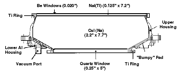
The crystals are contained in an opaque, hermetically sealed housing to prevent degradation of the NaI by water vapor and to shield the photomultiplier from stray light. The housing incorporates a 0.020-in thick beryllium x-ray entrance window to provide a light seal and a minimum of low energy photoelectric absorption of incident x-rays. The crystals are wrapped in teflon sheet, 0.010 in thick, and are highly polished to provide the maximum uniform light collection by the photomultiplier tube, and, therefore, maximize energy resolution. The photomultiplier tube and its attendant high voltage divider network (bleeder string), coupling elements, and connectors are surrounded by potting mate rial to meet vibration and thermal requirements, as well as to prevent electrical discharges at all ambi ent pressures. Ribbing moulded into the potting material provides a light seal and the compression properties to absorb thermal expansion and contraction without stressing the photomultiplier tube. The entire potted assembly is contained within a metallic housing that also acts as a magnetic shield.